How do blood clots travel from venous thrombosis to pulmonary embolism
Feb 14, 2026
Author: Wei Yun, Chief Nurse of the Vascular Surgery Department at Shanghai First People's Hospital. Review: Zhang Zhiwei, Deputy Chief Physician of the Vascular Surgery Department at Shanghai First People's Hospital. Meng Qingyou, Chief Physician of the Vascular Surgery Department at Shanghai First People's Hospital.
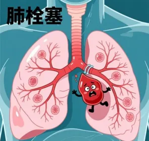
Title: How do blood clots travel from venous thrombosis to pulmonary embolism Introduction: Blood clots can lead to serious health issues like pulmonary embolism. This article explains thrombus formation and prevention measures. Keywords: ['Cardiovascular health', 'Prevention'] Main text: When you suddenly experience leg swelling or pain after sitting for a long time, or when your calves become stiff after lying in bed post-surgery, these seemingly ordinary symptoms could be the precursor to a life-threatening crisis—deep vein thrombosis in the lower limbs. This 'time bomb' lurking in the veins can detach and travel through the bloodstream to the lungs, instantly transforming into a pulmonary embolism, which can cause difficulty breathing, chest pain, hemoptysis, and even sudden death. Today, we will trace a thrombus that has 'run away' from the deep veins of the leg, embarking on a thrilling 'fantasy drift,' and observe how it gradually evolves into a potentially fatal 'pulmonary embolism.' First Stop: The Quiet Birth—Uninvited Guests in the 'Deep Veins'. Our story begins in the 'deep veins' of the lower limbs. Under normal circumstances, blood flows smoothly here. However, in some cases, blood clots can silently form in this tranquil passage, a condition medically known as 'deep vein thrombosis.' How is deep vein thrombosis formed? It requires three conditions, which are medically referred to as 'triad of thrombosis,' also known as 'Virchow's triad,' consisting of: 1. Venous blood flow stasis: For example, prolonged bed rest, long periods of sitting (such as during long flights or extended car journeys), paralysis, or reduced activity resulting from surgery or pregnancy. 2. Vascular endothelial injury: Damage occurs on the inner wall of blood vessels, making it prone to thrombosis. This often arises from severe trauma, major orthopedic surgeries, venipuncture catheterization, long-term smoking, and chemotherapy patients, among others. 3. Hypercoagulable state: This is commonly seen in older age (with increased risk for those over 60), obesity, during pregnancy and within six weeks postpartum, the use of certain medications (such as estrogen-based contraceptives), malignant tumors, blood disorders, and others. When these three 'pranks' come together, platelets and fibrin gather in the veins, accumulating like a snowball to form a solid mass—a thrombus. Initially, it may begin as a small 'sludge' near the venous valves, but it will gradually enlarge and block the blood vessel. At this point, the patient may experience swelling, pain, increased skin temperature, and redness or discoloration in the affected leg. However, the frightening aspect is that about half of deep vein thromboses show almost no obvious symptoms in their early stages, resembling a hidden bomb. Second stop: Thrilling drop – embarking on the 'rafting' journey. Recently formed blood clots, especially fresh ones, do not cling firmly to the walls of blood vessels. They resemble a clump of mud on a riverbed, and a careless 'rush of water' could easily dislodge them from their original position. What directly induces the dislodgment of blood clots? 1. External forces or changes in body position: Sudden movements after prolonged immobility, such as abruptly standing up or walking after sitting for a long time, severe coughing, injuries, or massage, may directly cause a thrombus to detach from the vessel wall. 2. Blood Flow Surge: When blood flow velocity suddenly increases (for example, during intense exercise), a thrombus may be 'washed away' by the blood flow, and the pressure generated by heart contractions or vascular pulsations may also increase the risk of thrombus dislodgment. 3. The instability of the thrombus itself: Fresh thrombi have a short formation time (ranging from several hours to a few days), and they have a softer texture, not tightly adhering to the vessel wall, making them prone to detachment. Parts of the partially dissolved thrombus have a loose structure, making them more easily carried away by blood flow. Third Stop: Fatal Journey - Breaking into the 'Lung Maze.' Once it falls off, this blood clot fragment no longer remains at rest. It is carried by the rushing venous blood and begins a 'drift' downstream. Its journey follows progressively larger veins, from the deep veins of the lower limbs to the iliac vein, then the inferior vena cava, the heart (right atrium and right ventricle), and finally to the pulmonary artery. The pulmonary artery and its branches resemble an inverted tree, with a thick trunk and increasingly finer branches. At this point, the clot's 'drift' encounters a bottleneck, acting like a large plug that suddenly blocks the blood flow to the lungs, leading to pulmonary embolism—a critical condition characterized by sudden onset and grave implications that can rapidly result in respiratory and circulatory failure. When the clot obstructs the pulmonary artery, it triggers a series of disastrous chain reactions. When a patient experiences a pulmonary embolism, what warning signs may manifest in the body? 1. Sudden shortness of breath: the most common and typical symptom, feeling 'out of breath,' with rapid, labored breathing. 2. Severe chest pain: It often occurs suddenly and intensifies with coughing, deep breathing, or bending over. 3. Hemoptysis: The patient may cough up phlegm with blood-streaks, and in severe cases, may cough up bright red blood. 4. Cough: A dry cough occasionally accompanied by a small amount of white mucus, with some patients experiencing wheezing. 5. Fainting or near-death sensation: Due to insufficient blood supply to the brain, the patient may suddenly faint and lose consciousness for a brief period. 6. In severe cases, sudden death can occur: A large blood clot blocking the main pulmonary artery can lead to death within minutes. Final Destination: The Storm Approaches—The Turbulence of Pulmonary Embolism. What should I do if a pulmonary embolism occurs? If deep vein thrombosis or pulmonary embolism is suspected, seeking immediate medical attention is the only option. Doctors will use D-dimer blood tests, lower limb venous ultrasound, and CT pulmonary angiography to make a diagnosis. 1. General treatment: Monitor the patient's condition changes, ensure absolute bed rest, and avoid strenuous activities and straining during bowel movements. 2. Anticoagulant therapy: Anticoagulant medications are used to prevent the further enlargement of thrombus and the formation of new thrombi. Commonly used medications include low molecular weight heparin, warfarin, and rivaroxaban. 3. Thrombolytic therapy: For massive pulmonary embolism that is life-threatening, thrombolytic agents can be injected directly into the pulmonary artery via intravenous or catheter methods to rapidly dissolve the thrombus. 4. Interventional therapy: thrombus aspiration and fragmentation through an intravascular catheter, or the placement of a filter in the inferior vena cava to intercept thrombi that have dislodged from the lower limbs. 5. Surgery: In extreme cases, perform a thoracotomy for pulmonary artery thromboectomy. How can we prevent deep vein thrombosis (DVT) in the lower limbs during daily life? 1. Reasonable Exercise: Get moving and avoid prolonged sitting or standing. During long trips, take regular breaks to stand up and stretch, flex your toes, and rotate your ankles, while ensuring you stay well-hydrated. Post-operative patients should get out of bed as early as possible under the guidance of their doctor. 2. Physical prevention: Wear medical compression stockings to promote venous blood return. Physical assistive devices such as intermittent pneumatic pressure devices or foot venous pumps may also be used. Elevating the legs at night can further enhance blood flow. 3. Standardize anticoagulation therapy: High-risk patients should use anticoagulants as prescribed by their healthcare provider, and must not stop or reduce the dosage on their own. It is essential to regularly monitor coagulation function during treatment and to be vigilant for signs of bleeding. 4. Healthy Lifestyle: Quit smoking, manage weight, and maintain a regular exercise routine to avoid staying in the same position for prolonged periods. Ensure a balanced diet, avoid irritating foods, keep bowel movements regular, and prevent increased abdominal pressure. From 'deep vein thrombosis' to 'pulmonary embolism,' it's not a whimsical adventure, but a critical emergency unfolding in the human body that demands urgency every second. Let us pay attention to vascular health, stay active, and remain vigilant.
#cardiovascular health
#prevention


© 2025 Health Tribe.